Beranda
/ Anatomy Of Ribs Posterior / 8 Muscles Of The Spine And Rib Cage Musculoskeletal Key - Posterior rib cage muscles :
Anatomy Of Ribs Posterior / 8 Muscles Of The Spine And Rib Cage Musculoskeletal Key - Posterior rib cage muscles :
Insurance Gas/Electricity Loans Mortgage Attorney Lawyer Donate Conference Call Degree Credit Treatment Software Classes Recovery Trading Rehab Hosting Transfer Cord Blood Claim compensation mesothelioma mesothelioma attorney Houston car accident lawyer moreno valley can you sue a doctor for wrong diagnosis doctorate in security top online doctoral programs in business educational leadership doctoral programs online car accident doctor atlanta car accident doctor atlanta accident attorney rancho Cucamonga truck accident attorney san Antonio ONLINE BUSINESS DEGREE PROGRAMS ACCREDITED online accredited psychology degree masters degree in human resources online public administration masters degree online bitcoin merchant account bitcoin merchant services compare car insurance auto insurance troy mi seo explanation digital marketing degree floridaseo company fitness showrooms stamfordct how to work more efficiently seowordpress tips meaning of seo what is an seo what does an seo do what seo stands for best seotips google seo advice seo steps, The secure cloud-based platform for smart service delivery. Safelink is used by legal, professional and financial services to protect sensitive information, accelerate business processes and increase productivity. Use Safelink to collaborate securely with clients, colleagues and external parties. Safelink has a menu of workspace types with advanced features for dispute resolution, running deals and customised client portal creation. All data is encrypted (at rest and in transit and you retain your own encryption keys. Our titan security framework ensures your data is secure and you even have the option to choose your own data location from Channel Islands, London (UK), Dublin (EU), Australia.
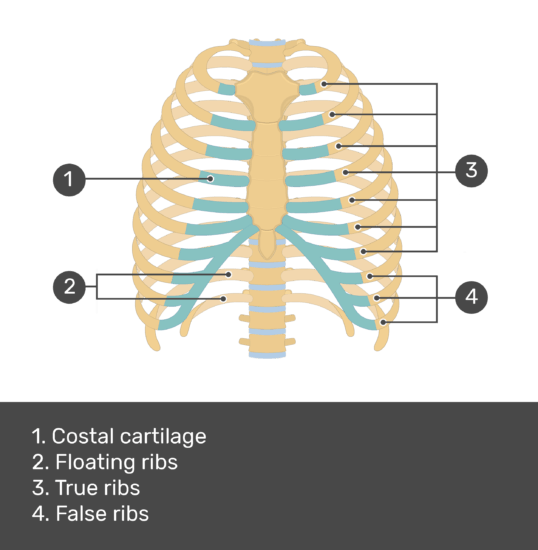
Anatomy Of Ribs Posterior / 8 Muscles Of The Spine And Rib Cage Musculoskeletal Key - Posterior rib cage muscles :. Anatomical anomalies at the thoracolumbar (t/l) and lumbocaudal (l/cd) boundaries of whales complicate the of vertebral articulation facets for posterior ribs and by ontogenetic rib fusion. Ribs eight to ten are the false ribs and are connected to the sternum indirectly via the cartilage of the rib above serratus posterior. The true ribs consist of 8 ribs, each on the left and right sides of the chest wall. Gross anatomy there are 12 pairs of ribs which are separated by intercostal spaces. This incision may be continued across the costal margin to open the abdominal cavity as in.
The posterior abdominal wall is a musculoskeletal structure formed by the posterior abdominal muscles, their fascia, the lumbar vertebrae and the image: Skeletal system anatomy and physiology nurseslabs. On anatomical parts the user can choose to display the various structures in colored illustrations of the anatomy of the back and spine: It is the area of articulation with the transverse process of the vertebra. Each rib forms two joints
Chest Wall Amboss from media-us.amboss.com The most superior rib is designated rib 1 and it articulates with the t1 thoracic vertebrae. Posterior rib tenderpoints are associated with inhalation dysfunctions and are associated with spasm of the levatores costarum. Made up of thoracic vertebrae, ribs and… functions at upper end to connect the shoulder girdle and conn… In vertebrate anatomy, ribs (latin: The nomenclature of the costal veins is the same as the arteries. Ribs 3 to 9 are considered typical ribs. Vertebrae, bones, joints, ligaments, muscles, muscular system, fascia, arteries, veins, nerves and various adjacent organs. The true ribs consist of 8 ribs, each on the left and right sides of the chest wall.
Each rib forms two joints
The lumbar plexus and its branches. by henry vandyke carter, henry gray (1918) anatomy of the human body. The most superior rib is designated rib 1 and it articulates with the t1 thoracic vertebrae. This incision may be continued across the costal margin to open the abdominal cavity as in. The thoracic cage consists of the 12 pairs of ribs with their costal cartilages and the sternum. There are twelve pairs of ribs. Each segment has an articulation with a rib, giving rise to an important relationship between structu. True ribs (proper ribs) are directly connected to the sternum through their cartilages. Major landmarks of a typical rib are the following: Includes images, video, and free quiz. Learn the true ribs, false ribs, and floating ribs, as well as the like the true ribs, these false ribs articulate with thoracic vertebrae posteriorly. 1.3 ribs anatomy and somatic dysfunctions. Each rib articulates posteriorly with two thoracic vertebrae by the costovertebral joint. In vertebrate anatomy, ribs (latin:
The most superior rib is designated rib 1 and it articulates with the t1 thoracic vertebrae. Skeletal system anatomy and physiology nurseslabs. This muscle is present posteriorly within the thoracic wall. The nomenclature of the costal veins is the same as the arteries. The thoracic spine, composed of 12 segments, is the longest subsection of the vertebral column.
Thoracic Skeleton Posterior View Stock Image Image Of Chest Skeletal 78412299 from thumbs.dreamstime.com The first seven pairs of ribs are true ribs as they are attached to the sternum directly by costal cartilages scalenus anterior, posterior and medius muscles have attachments on the first and second ribs. 1.3 ribs anatomy and somatic dysfunctions. The ribs are a set of twelve paired bones which form the protective 'cage' of the thorax. Head, neck, tubercle, and body of a rib. The ribs stretches posteriorly from thoracic vertebrae to the anterior lateral edges of the sternum. Vertebrae, bones, joints, ligaments, muscles, muscular system, fascia, arteries, veins, nerves and various adjacent organs. In vertebrate anatomy, ribs (latin: Made up of thoracic vertebrae, ribs and… functions at upper end to connect the shoulder girdle and conn…
An exception to this rule is that the first rib articulates with the first 20° to the frontal plane, with the superior facets facing posterior and a little up and laterally and the inferior facets facing anteriorly, down, and medially.
Nevertheless, denitive criteria for identication of cervical and caudal vertebrae leaves ambiguity only. The cords of the brachial plexus leave the posterior cervical triangle and enter the axilla through the axillary inlet. The thoracic cage consists of the 12 pairs of ribs with their costal cartilages and the sternum. However, they do not attach directly to the sternum anteriorly, and instead, attach to the. Further details of its anatomical relations and muscle attachments can be found in its own section in this text. Includes images, video, and free quiz. Both muscles attach to various ribs and parts of the spine. On anatomical parts the user can choose to display the various structures in colored illustrations of the anatomy of the back and spine: Each rib articulates posteriorly with two thoracic vertebrae by the costovertebral joint. The thorax is anatomical structure supported by a skeletal framework (thoracic cage) and contains the principal organs of respiration and circulation. The most superior rib is designated rib 1 and it articulates with the t1 thoracic vertebrae. This muscle is present posteriorly within the thoracic wall. Each rib forms two joints
In the anatomical position, the scapula overlies the second to seventh ribs on the posterolateral aspect of the chest wall. Skeletal system anatomy and physiology nurseslabs. Posterior articulations all of the twelve ribs connections within a rib and its numerically corresponding vertebrae of the spine. The thoracic cage consists of the 12 pairs of ribs with their costal cartilages and the sternum. Head, neck, tubercle, and body of a rib.
Structure Of The Ribcage And Ribs from www.getbodysmart.com Common characteristics of the ribs figs. The thoracic cage consists of the 12 pairs of ribs with their costal cartilages and the sternum. Each rib articulates posteriorly with two thoracic vertebrae by the costovertebral joint. Includes images, video, and free quiz. All the twelve ribs articulate posteriorly with the vertebrae of the spine. Anatomical anomalies at the thoracolumbar (t/l) and lumbocaudal (l/cd) boundaries of whales complicate the of vertebral articulation facets for posterior ribs and by ontogenetic rib fusion. On anatomical parts the user can choose to display the various structures in colored illustrations of the anatomy of the back and spine: The nomenclature of the costal veins is the same as the arteries.
Vertebrae, bones, joints, ligaments, muscles, muscular system, fascia, arteries, veins, nerves and various adjacent organs.
Be sure to subscribe to the visible body blog for more anatomy awesomeness! The most superior rib is designated rib 1 and it articulates with the t1 thoracic vertebrae. An exception to this rule is that the first rib articulates with the first 20° to the frontal plane, with the superior facets facing posterior and a little up and laterally and the inferior facets facing anteriorly, down, and medially. Each rib forms two joints Major landmarks of a typical rib are the following: On anatomical parts the user can choose to display the various structures in colored illustrations of the anatomy of the back and spine: Nevertheless, denitive criteria for identication of cervical and caudal vertebrae leaves ambiguity only. The posterior end is composed of head, neck, and tubercle. In the anatomical position, the scapula overlies the second to seventh ribs on the posterolateral aspect of the chest wall. The nomenclature of the costal veins is the same as the arteries. The thoracic spine, composed of 12 segments, is the longest subsection of the vertebral column. Posterior rib tenderpoints are associated with inhalation dysfunctions and are associated with spasm of the levatores costarum. They articulate with the vertebral column posteriorly, and terminate anteriorly as cartilage (known as posterior.
In most tetrapods, ribs surround the chest, enabling the lungs to expand and thus facilitate breathing by expanding the chest cavity anatomy of ribs. 1.3 ribs anatomy and somatic dysfunctions.